The MEIAF is a state-of-the-art imaging facility for both academic and non-academic research. It features an environmental scanning electron microscope (ESEM) with X-ray microanalysis and a cold stage that together enable high-resolution imaging of hydrated specimens, observation of dynamic experiments such as crystal formation and dehydration, freeze-fracturing, and ultra-low temperature imaging. Applications range from microelectronics to forensic science. MEIAF comprises equipment and personnel, which together offer a wealth of new imaging possibilities for researchers in academia, government, and industry.
Facility
MEIAF's environmental scanning electron microscope (ESEM) allows specimens to be maintained at near-ambient pressure during high-resolution imaging. This capability facilitates direct observation of dynamic experiments, such as crystal formation, hydration, and dehydration. The energy-dispersive spectrometer (EDS) enables X-ray detection and quantification for elemental analysis. A cryo stage and transfer system allows for frozen specimen observation and freeze-fracture preparation. MEIAF is staffed with full-time personnel who assist researchers with imaging and developing applications.
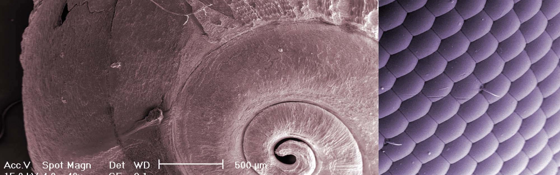
Many samples - including environmental, biological, and materials science specimens - are destroyed or masked by conventional scanning electron microscopy (SEM) preparation and imaging methods. The technology in MEIAF allows for conventional SEM but features environmental SEM (ESEM). ESEM circumvents destructive sample preparations and allows the option of imaging under a moderate vacuum and in a moist atmosphere. The result is that specimens are imaged in their native state. All specimens - including hard matter and even soft materials that are wet, oily, or frozen - are candidates for ESEM.
Technology
The core technology is an FEI Co. XL30 ESEM with a field emission gun (FEG). The ESEM detectors include a patented gaseous secondary electron detector (GSED), a solid-state backscattered electron detector (BSED), a large field detector (LFD), and a STEM detector for scanning transmission microscopy of embedded thin sections.
The ESEM can be used as a conventional SEM (high vacuum mode) or as an environmental SEM (wet mode, i.e. moderate vacuum and moist atmosphere). Wet mode is facilitated by flooding the sample chamber with water vapor and controlling condensation of the specimen surface via a temperature-controlled Peltier stage. Charging problems, common in materials with low electrical conductivity, do not occur because the water vapor neutralizes any charge that builds up on sample surfaces. Because sample drying and coating with a conductant are not necessary for wet mode, specimen preparation time is reduced, and biological samples and combined soft/hard matter can be imaged in their native state.
X-ray Microanalysis
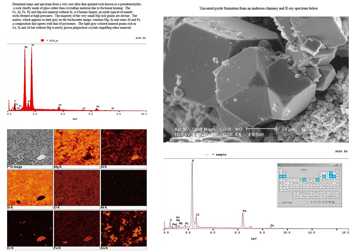
The ESEM is equipped with a Bruker energy-dispersive spectrometer (EDS) for X-ray microanalysis. The EDS has a QUANTAX XFlash 6|60 silicon drift detector (SDD) with energy resolution < 129 eV (MnΚα, 100 000 cps). ESPRIT microanalysis software is used for X-ray acquisition, qualitative and quantitative analysis, line profiles, and digital element maps.
Cryo Stage Imaging
The Quorum Technologies' Polaron PP2000T cryo stage extends MEIAF's ESEM capabilities to freeze-fracturing and ultra-low temperature imaging. With the cryo system, we can freeze and etch specimens, and maintain frozen samples at low temperature while imaging.
Applications
ESEM imaging has a myriad of applications that span diverse fields:
Archaeology and Physical Anthropology: Observation and analysis of uncoated archaeological artifacts and bones. Potential users: Archaeologists, researchers in physical anthropology.
Art/Museum Conservation: Depositional studies, observations of object surface degradation, elemental analysis of pigments or other artists' materials. Potential users: Art curators, art preservationists, art historians.
Biology, including Botany and Entomology: Examination of fresh or preserved specimens of plant seedlings, insects, mycelia, pollen grains, etc. Potential users: Museums of natural history, biological collections, biological research centers.
Cement Science: Fracture surface morphology, elemental impurity analysis. Potential users: Cement industry, adhesive and sealant industry, construction consultants.
Environmental Sustainability: Studies of engineered nanoparticles in plants and associated with microbes. Observations of colonized microfibers, plastics, and cigarette filters from terrestrial and marine environments. Agglomerations of heterogenous combined soft/ hard matter settled from water columns. Soil biofilms under varying wetting/ drying regimes.
Fiber Technology: Study of the effects of wetting and drying on different types of fibers, natural or synthetic; examination of fiber contaminants at high magnification. Potential users: Textile industry, pulp and paper industries, manufacturers of fiber-based materials.
Forensic: Observation of explosive residue, including gun powder; fiber and hair analysis. Potential users: Coroners, police departments, investigative units.
Geological Sciences/Petroleum Geology: Morphology of geological material such as microfossils, sediments, and mineral grains in their natural state, dry or wet. Potential users: Geologists, geochemical industry, oil, gas, and mineral extraction industries.
Hydrology, including Snow Science: Studies of snow sintering and disaggregation in real time; chemical characterization of impurities. Potential users: Snow hydrologists, resort managers, resource managers.
Materials Science: Study of polymers, ceramics (and other semiconductors that do not need to be coated to be observed), catalysts, food structure and properties, biomaterials, paper. Potential users: Polymer scientists, researchers in ceramic materials, food scientists, biomedical materials researchers and manufacturers, paper and paper product scientists and engineers.
Medicine: Observation of fresh and wet tissue. Potential users: Medical foundations that conduct research, research hospitals, schools of medicine.
Microbiology: Observation of bacterial growth on different types of substrate, both natural and produced in the lab. Potential users: Medical clinicians, food scientists, microbial ecologists, public health practitioners.
Microelectronics: Observation of integrated circuits, microsensors, microactuators and mechanical elements of MEMs. Potential users: Microelectronic and micromachining technology industries.
Pharmaceutical: Study of the dynamic physicochemical properties of drugs, such as swelling, dissolution, or disintegration, to understand the mechanisms of drug release. Potential users: Pharmaceutical companies with R & D departments.
Soil Science: Observation of untreated and treated soil samples at a magnification not previously possible. Potential users: Soil scientists, environmental consultants, soil microbiologists.
Rates
MEIAF equipment and services are available for the following rates (2018/2019):
- SEM usage (equipment only, internal use) - $93.22/hour
- SEM usage with Engineer (internal use) - $149.78/hour
- SEM usage (equipment only, off-campus use) - $144.49/hour
- SEM usage with Engineer (off-campus use) - $232.15/hour
This material is based upon work supported by the National Science Foundation under Awards BES-9977772, DBI-0216480, DEB-0444712, and by the National Science Foundation and the U.S. Environmental Protection Agency under DBI-0830117. Any opinions, findings, and conclusions or recommendations expressed in this material do not necessarily reflect the views of either the National Science Foundation or the Environmental Protection Agency. This material has not been subjected to Environmental Protection Agency review and no official endorsement should be inferred.
Contact
The MEIAF is open by appointment. Contact meiaf@bren.ucsb.edu with requests and questions.
Location:
Bren School of Environmental Science & Management
1006 Bren Hall, UCSB
Santa Barbara, CA 93106-5131